ECG & Pharmacology Online Course
- Description
- Curriculum
- FAQ
- Notice
- Reviews
ECG and Pharmacology Online Course – Essential Prep for ACLS & PALS Certification
Looking to strengthen your understanding of ECG interpretation and emergency pharmacology before taking ACLS or PALS training? Our ECG and Pharmacology Online Course is designed to give healthcare professionals a strong foundation in cardiac rhythm recognition and drug administration—key skills required before attending Advanced Cardiac Life Support (ACLS) or Pediatric Advanced Life Support (PALS) courses.
Since ECG and pharmacology are no longer covered in detail in ACLS and PALS certification courses, this online course ensures you’re fully prepared and confident when responding to cardiac emergencies.
✅ What You’ll Learn:
ECG Training:
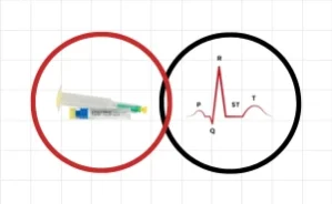
- Anatomy of the heart and cardiac conduction system
- Basics of electrophysiology
- How to measure and interpret a standard ECG
- Identification of basic arrhythmias
Emergency Pharmacology:
- Overview of core ACLS drugs and their clinical use
- Proper drug dosages and routes of administration in cardiac arrest and other emergencies
- How to integrate pharmacology into ACLS treatment algorithms
Whether you’re a new healthcare graduate or an experienced medical provider, this course will enhance your clinical skills and improve your readiness in high-stakes cardiac situations.
⚠️ Please note: This is not a Heart & Stroke Foundation course. It is intended as a preparatory course for ACLS and PALS training.
Updated on Jan 2025
-
1
 Before we start
 -
2
Welcome In our ECG & Pharmacology Training
the training will allow you to read and understand basic ECG
-
3
Anatomy of the Heart
Anatomy of the Heart” takes you on an illustrated journey through the structure and function of the human heart. With clear visuals and concise explanations, the video shows how blood flows through the atria and ventricles, how the valves keep everything moving in the right direction, and how the heart supports our entire circulatory system. A strong primer for anyone seeking to understand how this vital organ works — from its major chambers to its everyday role in keeping us alive.
-
4
The Heart
The video provides a concise description of the adult human heart's size and anatomical location:
-
Size: The heart of an adult is described as being "not much larger than a fist".
-
Location: It lies in the center of the chest.
-
It is positioned behind the breast bone, known as the sternum.
-
It is in front of the backbone, specifically the thoracic spine.
-
It is situated above the diaphragm.
-
-
Surroundings: Except for the area against the spine and a small strip down the center of the front, the heart is completely surrounded by the lungs.
-
-
5
The heart has its own blood supply
Coronary Arteries
The heart's independent blood supply system
-
6
The heart is a hollow organ
The heart is as a hollow organ divided into four sections or chambers
-
7
Conduction of the Heart 1
The video focuses on the initial components and pathways involved in the heart's electrical conduction system:
-
8
Conduction of the Heart (2)
This video details the structure and function of the Atrioventricular (AV) node and its connection to the rest of the heart's electrical system.
-
9
Conduction of the Heart (3)
This video details the structure and paths of the heart's bundle branches.
Left Bundle Branch
Right Bundle Branch
-
10
Conduction of the Heart (4)
This video describes the path and speed of the electrical impulse as it travels through the heart's lower chambers, the ventricles.
-
11
ECG Components
This video defines the major waves and intervals found on an Electrocardiogram (ECG)
-
12
Rhythm indicators
Electrical Events and ECG Components
the major electrical events occurring in the heart with their corresponding segments and waves on an ECG:
-
13
ECG Measurement 1
The standard speed and time measurements associated with ECG paper:
-
14
ECG Measurement 2
Details the measurements on an ECG grid and the normal durations for key ECG intervals:
-
ECG Grid Axes:
-
The horizontal axis measures time.
-
The vertical axis measures voltage or amplitude.
-
1 millivolt (mV) is equal to two large boxes high.
-
-
Normal ECG Interval Measurements:
-
-
15
Heart Rate Estimation
Heart Rate Estimation Methods
outlines several methods for estimating heart rate from an ECG strip, noting that a simpler method is often used at the bedside: -
16
Lead Placement
Monitoring Leads vs. Diagnostic ECG
clarifies the difference between monitoring leads (like standard monitoring in Lead II) and a 12-lead diagnostic ECG:
-
17
Rhythm Analysis
Four Questions for Rhythm Interpretation
This standard, consistent, and routine approach to rhythm analysis is key to success. It presents four basic questions that lead to the correct analysis and diagnosis of most rhythm abnormalities:
-
18
Normal Sinus Rhythm
Criteria for Normal Sinus Rhythm (NSR)
This is list of key characteristics used to identify Normal Sinus Rhythm on an ECG:
-
19
Sinus Bradycardia
Criteria for Sinus Bradycardia
This is key characteristics used to identify Sinus Bradycardia on an ECG:
-
20
Sinus Tachycardia
-
21
Premature Atrial Contraction (Complex)
Criteria for Premature Atrial Contraction (PAC)
The key characteristics used to identify a Premature Atrial Contraction (PAC) complex on an ECG:
-
22
Supraventricular Tachycardia
Criteria for Supraventricular Tachycardia (SVT)
This is the key characteristics used to identify Supraventricular Tachycardia (SVT) on an ECG:
-
23
Premature Ventricular Contraction (Complex)
Criteria for Premature Ventricular Contraction (PVC)
The lists the key characteristics used to identify a Premature Ventricular Contraction (PVC) complex on an ECG:
-
24
Monomorphic Ventricular Tachycardia
Criteria for Monomorphic Ventricular Tachycardia (V-Tach)
key characteristics used to identify Monomorphic Ventricular Tachycardia on an ECG:
-
25
Polymorphic Ventricular Tachycardia
Video Content Summary: Criteria for Polymorphic Ventricular Tachycardia (V-Tach)
The key characteristics used to identify Polymorphic Ventricular Tachycardia on an ECG:
-
26
Ventricular Fibrillation
Criteria for Ventricular Fibrillation (V-Fib)
There is a very concise description of the ECG characteristics of Ventricular Fibrillation:
-
Key Finding: When analyzing Ventricular Fibrillation, the video states that everything is absent. This refers to the absence of discernible P waves, QRS complexes, T waves, and a measurable rhythm or rate.
-
-
27
Asystole
Asystole on an ECG.
-
Asystole is characterized by an absence of any electrical activity, often referred to as a "flatline," meaning there are no discernible P waves, QRS complexes, or T waves.
-
-
28
Pulseless Electrical Activity
Criteria for Pulseless Electrical Activity (PEA)
This video explains the defining feature and variable ECG characteristics of Pulseless Electrical Activity (PEA):
-
29
First-Degree Block
Criteria for First Degree Block
This video lists the key characteristics used to identify a First Degree Atrioventricular (AV) Block on an ECG:
-
30
Second-Degree Block (Type I)
Criteria for Second Degree Block Type I (Wenckebach)
This is the key characteristics used to identify a Second Degree Atrioventricular (AV) Block Type I (also known as Mobitz I or Wenckebach) on an ECG:
-
31
Second-Degree Block (Type II)
Criteria for Second Degree Block Type II (Mobitz II)
the key characteristics used to identify a Second Degree Atrioventricular (AV) Block Type II (also known as Mobitz II) on an ECG:
-
32
Third-Degree Block
Criteria for Third Degree Block (Complete Heart Block)
This is lists the key characteristics used to identify a Third Degree Atrioventricular (AV) Block, also known as Complete Heart Block, on an ECG:
-
33
 ECG Quiz
-
34
Pharmacology Lesson
Pharmacological Tools
This video serves as a brief introduction to the use of pharmacological tools within an Advanced Cardiovascular Life Support (ACLS) context:
-
The video discusses the common drugs used in ACLS and directs the viewer to Table 1 in their corresponding ACLS manual for detailed information, including doses, routes, and uses.
-
Important Disclaimer: The video emphasizes that the use of any ACLS medication must be done within the provider's scope of practice and only after a thorough study of the drugs' actions and side effects.
-
Table 1 is intended only as a brief reminder for those who are already knowledgeable about these medications.
-
It specifically contains only adult doses, indications, and routes of administration for the most common ACLS drugs.
-
-
35
Pharmacology Test
Registered Nurses (RNs)
Nurse Practitioners (NPs)
Paramedics
Students in these fields
❓ Course Overview FAQs
1. What is the primary purpose of this course?
This course is an essential preparatory course for healthcare professionals planning to take the Advanced Cardiac Life Support (ACLS) or Pediatric Advanced Life Support (PALS) certification. It provides a strong foundation in ECG interpretation and emergency pharmacology, which are no longer covered in detail during the main ACLS and PALS courses.
2. Who is the intended audience for this course?
The course is designed for healthcare professionals, including:
Registered Nurses (RNs)
Nurse Practitioners (NPs)
Paramedics
Students in these fields
3. What topics are covered in the ECG training section?
The ECG portion of the course covers:
Anatomy of the heart and the cardiac conduction system.
Basics of electrophysiology.
How to measure and interpret a standard ECG.
Identification and criteria for basic arrhythmias, such as Normal Sinus Rhythm, Sinus Bradycardia, Sinus Tachycardia, Supraventricular Tachycardia (SVT), Ventricular Fibrillation (V-Fib), Asystole, and Pulseless Electrical Activity (PEA).
An overview of core ACLS drugs and their clinical use.
Proper drug dosages and routes of administration in cardiac emergencies.
How to integrate pharmacology into ACLS treatment algorithms.
⚠️ Please note: This is not a Heart & Stroke Foundation course. It is intended as a preparatory course for ACLS and PALS training. Updated on Jan 2025
Computer with Internet access
- RNs
- NPs
- Paramedics
- Students